
Finite Element Simulations

Throughout their life, most types of living cells are subjected to various mechanical forces which play a significant role in development of the cells. Mechanical behavior of the living cells is very important for understanding cells' biological processes such as development, differentiation, disease etc.

In order to investigate elastic and viscoelastic behavior of cells we use Finite Element Analysis (FEA) using software MSC Marc Mentat. In particular, we modeled cells' behavior under loading experienced during two experimental techniques: atomic force microscopy (AFM) and micropipette aspiration.


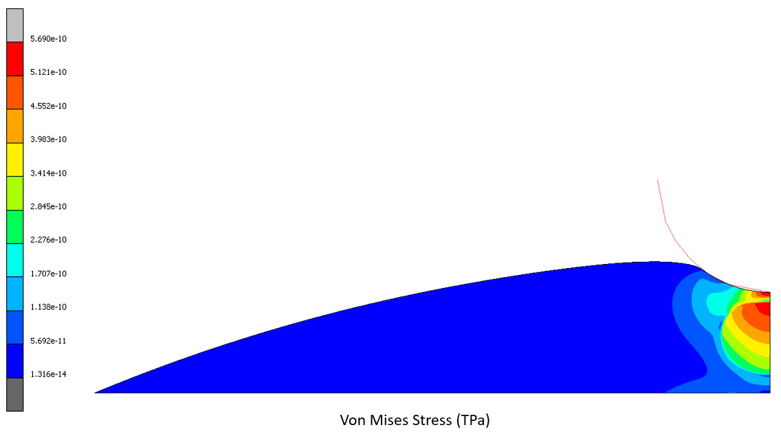
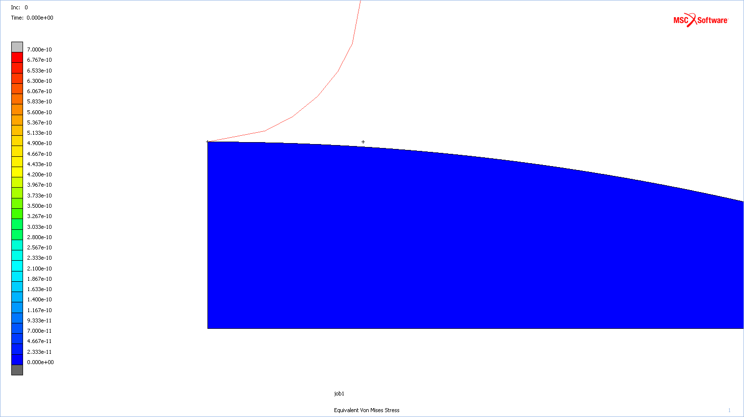
Axisymmetric FEA simulation of nanoindentation of a cell on a substrate

Nanoindentation FEA -- model


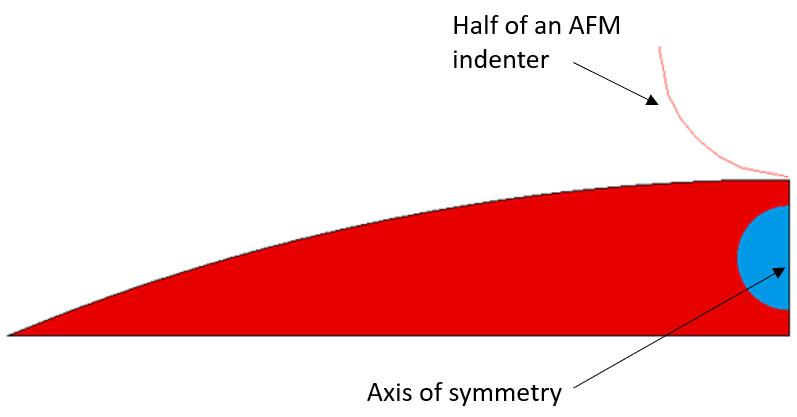
Using atomic force microscopy (AFM) a cell is subjected to indentation on nanoscale and as a result force-displacement curve can be obtained. In this FEA simulation we modeled a cell body (red) and a nucleus (blue). No other internal cell components were modeled. Both cell body and nucleus are described as elastic materials with Young's modulus and Poisson's ratio. The constant force is applied to the center of the cell.

Links to additional images:


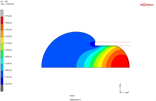
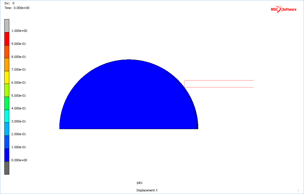
Axisymmetric FEA simulation of micropipette aspiration of a cell

Micropipette Aspriation FEA -- model


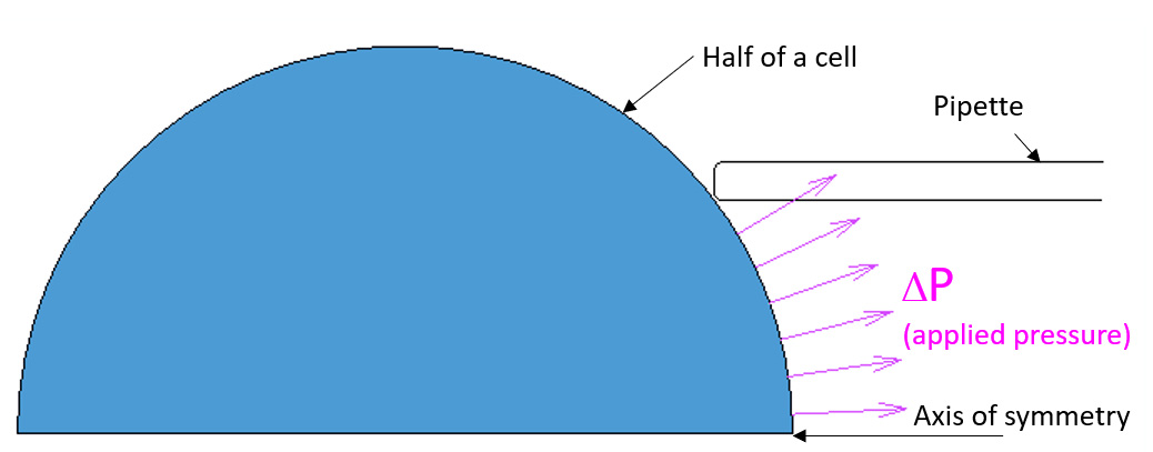
The micropipette aspiration (MA) is a non-invasive experimental technique which is used to study time-dependent deformation of the cells subjected to extracellular pressure ΔP.
In this FE simulation the cell is modeled as aspherical and homogeneous without specifying internal structure. Prony series were used to describe viscoelastic properties of the cell.

Links to additional images:

Membrane Building

Membrane building -- flowchart

The web-based graphical user interface Chemistry at Harvard Macromolecular Mechanics (CHARMM) was used to create a simple membrane and set parameters for running simulations. The protocol on making a single lipid membrane was to parameterize the membrane and choose the size of the system (200 x 200 Å). The lipid bilayer was generated with surrounding water solvent. These components required assembly and defining initial parameters to maintain a constant pressure and temperature along with calculating the present intermolecular and intramolecular interactions. The force field and the grid information was generated during the equilibration run. With the output files the simulation was run using the generated Groningen Machine for Chemical Simulations (Gromacs) input

Membrane building -- forces

Molecular Dynamics Simulations

Molecular dynamics simulations -- flowchart

Molecular dynamics is the study of molecular movement. The global motions are observed using the Coarse-Grained (CG) method which pairs 3-4 atoms into one point. This requires the use of a MARTINI Force-Field when calculating interactions with other particles. The goal is to predict the position of the molecules while acknowledging the intermolecular and intramolecular interactions occurring. This method saves computational time and resources and provides satisfactory data to collect mechanical values.

Newtons equation of motion (find trajectory of particles)

  • Total force for each particle is determined based on its interactions with other particles (Force-Field)

Molecular dynamics simulations -- forces

Visualization

Visualization -- left
Visualization -- right

The output files of the production run (structure .gro file and trajectory file .xtc) were input to the Visualization Molecular Dynamics software (VMD). The full simulation was observed to ensure structural errors were not present. The analysis of the fluctuation spectra required the membrane surface data which used the coordinates of the phosphorus atoms.